Saturday, 30 November 2013

Central Serous Retinopathy: CSR

WHAT IS CSR?

Central serous retinopathy (CSR) is a condition of unknown cause in which serous fluid ( thin and serum like fluid ) accumulates underneath the central part of the retina ( light sensitive part of the eye ). This fluid collection is similar to blisters caused on the skin when exposed to boiling water. This fluid collection beneath it causes the retina to become elevated. The elevation produces a change in the layout of the light sensors in the eye leading onto distorted and blurred vision.

WHO GETS CSR?

Most patients with CSR are males in their third and fourth decades. Stress or corticosteroid use may play roles in inciting or aggravation the condition. Intake of systemic steroids for Asthma and steroid containing ointments for skin disorders can also predispose to CSR. Hypertension and Caffeine use has also been implicated in CSR.

CSR is more common in anxious and stressed out persons. It can occur after a period of intense stress like divorce or loss of job.

WHAT ARE THE SYMPTOMS OF CSR?

Many patients first notice a minor blurring of vision, followed by various degrees of:
  • metamorphopsia (defective, distorted vision)
  • micropsia (distorted visual perception in which objects appear smaller than their actual size)
  • chromatopsia (visual defect in which objects appear unnaturally colored)
  • central scotoma
  • increasing hyperopia (farsightedness)
Visual acuity in the acute stage may range from 20/20 to 20/200 and averages 20/30. In some patients the onset of symptoms is preceded or accompanied by migraine-like headaches.


WHAT IS THE NATURAL COURSE OF CSR?

When left alone, central serous retinopathy heals spontaneously within 4 to 8 weeks, with full recovery of visual acuity. However, about one-third to one-half of all patients have recurrences after the first episode of the disease; 10 percent have three or more recurrences. In almost half of the patients, the recurrence is within one year of the primary episode, but relapses may occur up to ten years later.

Central serous choroidopathy usually affects just one eye at a time, but it is possible that both eyes may be affected at the same time.


WHAT TESTS MUST BE DONE?

The diagnosis is made by an eye examination, sometimes using a fundus contact lens. The diagnosis is confirmed by fluorescein angiography. In this test, an orange dye is injected into the patient’s vein, and this dye is observed as it circulates through the ocular vasculature. Typically the fluorescein enters into the blister and stains its contents, identifying one or more leakage points. Often, areas of previous retinal pigment epithelial disruption can be visualized elsewhere in the same eye or in the macula of the unaffected eye. Ocular coherence tomography (OCT) is another test that is helpful. This imaging modality can accurately detect fluid and swelling in the retina.


HOW DO WE TREAT CSR?            

The typical patient with central serous retinopathy does not require treatment. Most episodes of this condition are self-limited and resolve within 2 to 3 months. Simple reassurance to the patient that this will occur is beneficial. In some patients, the subretinal fluid persists for longer than this time period, and, in this setting, treatment is recommended. Thermal laser can be applied to the area of the retinal pigment epithelium that is thought to be playing a role in fluid accumulation.  Usually, this treatment is quite effective and can be very useful when the area of leakage is well away from the center of vision.  Many patients will recover vision back to the 6/6 level, however, patients will often report that vision in the affected eye is not quite as good as the normal eye even long after the fluid has resolved. Approximately 25% of patients develop a recurrent episode. There is also a subset of patients with significant retinal pigment epithelial abnormalities that are predisposed to attacks in both eyes, often simultaneously, and the prognosis in this group is much less optimistic, though with treatment many can maintain reading and driving vision.

WHAT CAN YOU DO TO PREVENT RECURRENCE?

If you have had an attack of CSR you have a high chance of having a recurrence. To lower the risk of recurrence:

  • ·         Learn relaxation techniques like yoga and meditation.
  • ·         Try to find solution to constant stressors in life
  • ·         Exercise daily
  • ·         Totally abstain from smoking
  • ·         Loose body weight if you are over weight


Monday, 14 October 2013

TAKING CARE OF YOUR CHILD’S EYES

We all know how precious eyes are and to make sure our children enjoy good vision throughout their life time we need to ensure that they receive proper eye care. If found early, many  eye conditions can be effectively treated. Sometimes delay in treatment can mean permanent loss of vision for the child.

Eye Exams
Routine medical exams for kids' vision include:
  • Newborns should be checked for general eye health by a pediatrician or family physician in the hospital nursery.
  • High-risk newborns (including premature infants), those with a family history of eye problems, and those with obvious eye irregularities should be examined by an eye doctor.
  • In the first year of life, all infants should be routinely screened for eye health during checkups with their pediatrician or family doctor.
  •  Around age 5, kids should have their vision and eye alignment evaluated by an Ophthalmologist.
  • After age 5, further routine screenings should be done if the child complains of blurred vision, squinting or frequent headaches
  •   Kids who wear prescription glasses or contacts should have annual checkups by an eye doctor to screen for vision changes.

Spotting Eye Problems
Signs that a child may have vision problems include:
  • constant eye rubbing
  • extreme light sensitivity
  • poor focusing
  • poor visual tracking (following an object)
  • abnormal alignment or movement of the eyes (after 6 months of age)
  • chronic redness of the eyes
  • chronic tearing of the eyes
  • a white pupil instead of black
In school-age children, watch for other signs such as:
  • inability to see objects at a distance
  • inability to read the blackboard
  • squinting
  •  difficulty reading
  •  sitting too close to the TV

Common Eye Problems
Several eye conditions can affect kids. Most are detected by a vision screening using an acuity chart.
·         Amblyopia ("lazy eye") is poor vision in an eye that may appear to be normal. Two common causes are crossed eyes and a difference in the refractive error between the two eyes. If untreated, amblyopia can cause irreversible visual loss in the affected eye. (By then, the brain's "programming" will ignore signals from that eye.)  Amblyopia is best treated during the preschool years.
·         Strabismus is a misalignment of the eyes; they may turn in, out, up, or down. If the same eye is chronically misaligned, amblyopia may also develop in that eye. With early detection, vision can be restored by patching the properly aligned eye, which forces the misaligned one to work. Surgery or specially designed glasses also may help the eyes to align.
·         Refractive errors mean that the shape of the eye doesn't refract, or bend, light properly, so images appear blurred. Refractive errors also can cause amblyopia. Nearsightedness is the most common refractive error in school-age children; others include farsightedness and astigmatism:
o    Nearsightedness is poor distance vision (also called myopia), which is usually treated with glasses or contacts.
o    Farsightedness is poor near vision (also called hyperopia), which is usually treated with glasses or contacts.
o    Astigmatism is imperfect curvature of the front surface of the eye, which is usually treated with glasses if it causes blurred vision or discomfort.
Other eye conditions require immediate attention, such as retinopathy of prematurity (a disease that affects the eyes of premature babies) and those associated with a family history, including:

·         Retinoblastoma is a malignant tumor that usually appears in the first 3 years of life. The affected eye or eyes may have visual loss and whiteness in the pupil.
·         Infantile cataracts can occur in newborns. A cataract is a clouding of the eye's lens.
·         Congenital glaucoma in infants is a rare condition that may be inherited. It is the result of incorrect or incomplete development of the eye drainage canals before birth and can be treated with medication and surgery.
·         Genetic or metabolic diseases of the eye, such as inherited disorders that make a child more likely to develop retinoblastoma or cataracts, may require kids to have eye exams at an early age and regular screenings.


Glasses and Contacts
Kids of all ages — even babies — can wear glasses and contacts.

Keep these tips in mind for kids who wear glasses:
  • Allow kids to pick their own frames.
  • Plastic frames are best for children younger than 2.
  •  If older kids wear metal frames, make sure they have spring hinges, which are more durable.
  • An elastic strap attached to the glasses will help keep them in place for active toddlers.
  • Kids with severe eye problems may need special lenses called high-index lenses, which are thinner and lighter than plastic lenses.
  • Polycarbonate lenses are recommended for all kids, especially for kids who play sports. Polycarbonate is a tough, shatter-proof, transparent thermoplastic used to make thin, light lenses. However, although they're very impact-resistant, these lenses scratch more easily than plastic lenses.
Infants born with congenital cataracts may need to have their cataracts surgically removed during the first few weeks of life. Some children born with cataracts wear contact lenses after cataract surgery.
Around age 10, kids may express a desire to get contact lenses for cosmetic reasons or convenience if they play sports. Allowing a child to wear contacts depends on his or her ability to insert and remove lenses properly, faithfully take them out as required, and clean them as recommended by the doctor. Contact lens problems are almost always caused by poor habits and bad hygiene.

Tuesday, 10 September 2013

Stye

What is a stye?
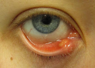
A stye is a tender, painful red bump located at the base of an eyelash or under or inside the eyelid. A stye results from a localized infection of the glands or a hair follicle of the eyelid. Scientifically a stye is called Hordeolum. It can be of two types: external hordeolum refers to a stye that develops at the base of an eyelash, whereas internal hordeolum refers to a stye that develops in a meibomian gland, a gland located on the underside of the eyelid that secretes an oily substance onto the eyeball.
When the infection of a stye persists over time, it can result  in scarring around the meibomian gland causing an usually painless hard swelling in the eyelid called Chalazion.


What is the cause of a stye?
A stye results from an infection of the oil glands of the eyelid that help to lubricate the eyeball. The infection occurs after these glands have become clogged. A stye also may arise from an infected hair follicle at the base of an eyelash. The bacterium Staphylococcus aureus that is frequently found on the skin is responsible for 90%-95% of cases of styes.

What are stye symptoms and signs?
The first signs and symptoms of a stye are usually redness, tenderness, and pain in the affected area. The eye may feel irritated or "scratchy." Later signs and symptoms may include swelling, discomfort during blinking of the eye, watering of the eye, and sensitivity to light. A common sign of a stye is a small, yellowish spot at the center of the bump that represents pus rising to the surface.

How is a stye diagnosed?
A stye is diagnosed by its characteristic appearance and symptoms. No other tests are necessary to establish the diagnosis of a stye.

Who is most susceptible to the development of a stye?
Styes are very common. People of all ages can develop a stye. Men and women are equally affected. People with certain chronic conditions like diabetes, seborrhea, and chronic debilitating illnesses are more prone to develop styes than the general population. In many susceptible people, stress seems to trigger the development of a stye. Studies have shown that those who have high levels of blood lipids are more susceptible to blockages in the oil glands, and are, therefore, more likely to develop a stye.
Persons with refratice error not using the correct eye glass power can get recurrent stye. Reccurent stye is also common in uncontrolled diabetics.

What is the treatment for a stye? Is there a home remedy for a stye?
Most styes will drain and resolve on their own without the need for medical treatment. Application of a warm compress to the affected area for 10-15 minutes, four to six times a day, can be an effective home remedy and speed rupture of the stye. This will aid in the relief of symptoms. A stye should not be pressed or squeezed since this can spread or worsen the infection. If a stye persists for several days, a doctor may incise  the swelling under local anesthesia in his or her office. Babies or children who require surgical drainage of a stye may need a general anesthetic. Antibiotic ointments are prescribed to treat a stye. Rarely, systemic (oral) antibiotics are recommended for persistent or multiple styes. Over-the-counter pain medications may be used to alleviate pain and tenderness. Contact lenses and eye makeup should never be worn during treatment for a stye.

What is the prognosis (outcome) of a stye?
A stye is harmless in the majority of cases. In most cases, a stye ruptures on its own within a few days to a week, leading to relief from symptoms. Some people will require medical or surgical treatment of a stye, as with complications described in the following section. A stye does not cause damage to the eye. Styes often recur, but complications of styes are rare.

Are there any potential complications resulting from a stye?
Complications of a stye are rare. The infection may spread to other eyelash follicles, leading to multiple styes. A chalazion is the most common complication that develops from a stye. Chalazia can be large enough to deform the cornea of the eye and interfere with vision, and they may cause a cosmetic problem. Other potential complications include spreading of the infection to the eyelids or other tissues near the eye. Improper drainage of a stye may lead to deformity or disruption of growth of eyelashes. Progression of a stye to a systemic infection (spreading throughout the body) is extremely rare, and only a few instances of such spread have been reported.

Can a stye be prevented?
While it is impossible to completely prevent the development of a stye, good hygienic practices, including proper hand washing, can help prevent all forms of infection, including a stye. Other measures that can help prevent styes include
  • ·         never sharing cosmetics or cosmetic tools such as lash curlers or eyelash combs with others,
  • ·         keeping cosmetic tools clean,
  • ·         discarding old or contaminated eye makeup,
  • ·         keeping all cosmetics clean,
  • ·         not touching the eye and surrounding areas.